A model trained similarly to ChatGPT has been created by researchers and may be modified to assess a variety of medical issues.
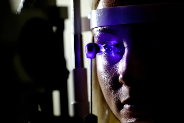
On the basis of a person’s retinal scans, researchers have created an artificial intelligence (AI) tool that can diagnose and foretell the likelihood of developing a variety of health disorders, such as Parkinson’s disease, heart failure, and eye ailments.
The new tool, named RETFound, stands out because it was created using a technique known as self-supervised learning. Previous AI programs have been trained to detect disease using retinal scans. Thus, it was unnecessary for the researchers to individually evaluate and categorize each of the 1.6 million retinal images utilized for training. The majority of common machine-learning models require the use of such time- and money-consuming processes.
Instead, the researchers employed a technique similar to that used to train ChatGPT, a large-language model. To learn how to guess the next word in a sentence based on the context of the preceding words, that AI program makes use of numerous examples of human-generated text. In a similar manner, RETFound learns how to forecast what missing areas of images should look like using a large number of retinal shots.
Pearse Keane, an ophthalmologist at Moorfields Eye Hospital NHS Foundation Trust in London and a co-author of a paper describing the tool that was published today in Nature1, claims that the model “somehow learns what a retina looks like and what all the features of a retina are” over the course of millions of images. This serves as the model’s cornerstone and qualifies it as what some refer to as a foundation model, meaning that it can be modified for a variety of jobs.
Providing insight into human health
The only area of the human body through which the capillary network, which is made up of the tiniest blood vessels, can be directly examined, the retinas, can provide a window into a person’s health. According to Keane, “we can directly visualize [that] in retinal images if you have some systemic cardiovascular disease, like hypertension, which is potentially affecting every blood vessel in your body.”
Retinal pictures can be utilized to assess neural tissue since they are both an extension of the central nervous system and resemble the brain. The problem is that most people lack the knowledge necessary to properly interpret these scans. Here’s where AI can help, according to Keane.
Keane and his colleagues could then introduce a small number of labelled images — say, 100 retinal images from people who had developed Parkinson’s and 100 from people who had not — to teach the model about particular conditions after they had pre-trained RETFound on those 1.6 million unlabeled retinal images. According to Keane, the model is able to easily understand the retinal traits connected to a disease because it has learned from all the unlabelled images what a retina should be like.
According to Xiaoxuan Liu, a clinical researcher at the University of Birmingham in the UK who researches responsible innovation in AI, using unlabelled data to first train the model “unblocks a major bottleneck for researchers.” Curtis Langlotz, a radiologist and the director of Stanford University’s Center for Artificial Intelligence in Medicine and Imaging, concurs. Because high-quality labels for medical data are so expensive, he claims that label efficiency has replaced quality as the gold standard.
When it came to recognizing eye conditions like diabetic retinopathy, the method worked effectively. Depending on the data set utilized, it scored between 0.822 and 0.943 for diabetic retinopathy on a scale where 1 corresponds to a perfect model that predicts accurately every time and 0.5 corresponds to a model that performs no better than a random prediction. The overall performance was limited when forecasting the risk for systemic disorders, such as heart attacks, heart failure, stroke, and Parkinson’s, but it was still better than previous AI models’ predictions in this area.
According to Liu, RETFound is one of the few foundation model applications for medical imaging that has so far been successful.
Expanding applications
In the future, researchers hope to apply the methods used to create RETFound to other types of medical imaging. “It will be interesting to see whether these methods generalize to more complex images,” adds Langlotz, citing magnetic resonance images and computed tomography scans as examples. These images are sometimes three- or even four-dimensional.
The model has been made accessible to the public in the authors’ hopes that organizations throughout the globe would be able to modify and train it to function for their local patient populations and healthcare environments. They might tweak this algorithm using data from their own nation to produce something that is better suited to their needs, says Keane.
This is really exciting, adds Liu. However, she adds, there is a risk involved in employing RETFound as the foundation for additional illness detection models. That’s because any restrictions incorporated into the tool could seep into derived models in the future. It is now up to the creators of RETFound to assure its moral and secure application, as well as open disclosure of its limitations, in order for it to truly serve the community.