According to a study, artificial intelligence is almost twice as effective as the current approach in evaluating the aggressiveness of a rare type of cancer through scans.
AI, with the ability to discern details that are imperceptible to the human eye, exhibited an 82% accuracy rate, in contrast to the 44% accuracy achieved through traditional lab analysis, as per findings. Researchers from the Royal Marsden Hospital and the Institute of Cancer Research are optimistic about its potential to enhance treatment and provide substantial benefits to thousands of patients annually.
Moreover, they are excited about the AI tool’s potential in early detection of other types of cancer. AI has already demonstrated significant promise in diagnosing breast cancer and reducing treatment durations. By processing vast amounts of data and learning to recognize patterns, AI systems can make predictions, solve complex problems, and even improve their performance through self-learning.
Professor Christina Messiou, a consultant radiologist at The Royal Marsden NHS Foundation Trust and a professor in imaging for personalized oncology at The Institute of Cancer Research, London, expressed great enthusiasm about the potential of this advanced technology, stating, “We’re incredibly excited by the potential of this state-of-the-art technology. It could lead to patients having better outcomes, through faster diagnosis and more effectively personalized treatment.”

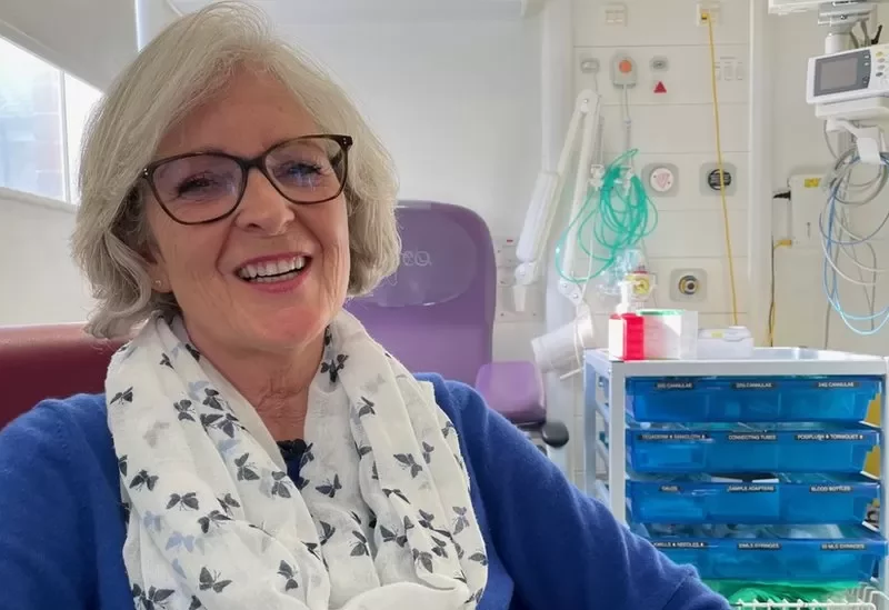
Tina had a sarcoma located in the posterior part of her abdomen.
In their study published in Lancet Oncology, the researchers employed a method known as radiomics to detect subtle indicators, imperceptible to the naked eye, of retroperitoneal sarcoma, a type of cancer that arises in the connective tissue at the rear of the abdomen. They analyzed scans from 170 patients with this technique.
Using the data obtained, the AI algorithm demonstrated a much higher accuracy in grading the aggressiveness of tumors in 89 additional patients from European and US hospitals based on scans, surpassing the precision of biopsies, which involve microscopic analysis of a small portion of cancerous tissue.
The AI Tool Allows for a Faster and More Efficient Diagnosis.
Tina McLaughlan, a dental nurse from Bedfordshire, received a sarcoma diagnosis in June of the previous year after experiencing stomach pain. Doctors used computerized tomography (CT) scan images to identify the issue and opted not to perform a needle biopsy due to the risks involved. Subsequently, they removed the tumor, and Tina now undergoes scans at the Royal Marsden every three months for follow-up.
While Tina wasn’t part of the AI trial, she believes it could be highly beneficial for other patients. She mentioned that initially, during the first scan, doctors couldn’t provide a definitive diagnosis. In her case, it wasn’t disclosed until after the histological analysis following the operation. Tina expressed her hope that the AI tool would lead to faster and more accurate diagnoses for patients in the future.
“Tailored Therapy”
Around 4,300 individuals in England receive a diagnosis of this cancer variety annually.
Professor Messiou envisions the possibility of implementing this technology globally, where high-risk patients can receive personalized treatment, while low-risk patients can avoid unnecessary treatments and subsequent scans.
Dr. Paul Huang, affiliated with the Institute of Cancer Research in London, expressed his optimism about the potential of this technology to revolutionize the lives of sarcoma patients by facilitating customized treatment strategies aligned with the unique characteristics of their cancer. He commended the promising results from this development.


